Neurons
The neuron (nerve cell) is the information-processing and information-transmitting element of the nervous system. Neurons come in many shapes and varieties depending on the specialised jobs they perform. Most neurons have, in one form or another, have three structures:
1) cell body
2) dendrites
3) axon
These are shown in the diagram above.
Dendrites are the part of the neuron which receives communications from another source i.e. information travelling from neuron to neuron. The cell body contains the nucleus and other vital organelles. Its shape varies considerably in different kinds of neurons. The axon is the part of the neuron which send the message/information (through the terminal buttons) elsewhere. The basic message which it carries is called an action potential. Talking physically, the axon is a long, thin tube which is often covered by a myelin sheath. This is shown in the diagram below (the blue layer covering the axon):
This diagram is demonstrating the principle parts of a multipolar neuron. This is the most common type of neuron found in the central nervous system.
Neurons are massively specialised and therefore need protection, fuel (sugars) and 'cleaning'.
Glial Cells
Glia are the most important supporting cells in the central nervous system. Glial cells have a 10:1 ratio to neurons. There are four types of Glial cell:
1) Oligodendrocytes
2) Schwann Cells
3) Astrocytes
4) Microglia
Glial cells wrap neurons in a protective layer (fatty sheath) and are drawn towards chemicals released by neurons.
Neural Conduction
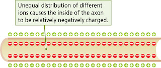
To measure the electrical charges generated by an axon, you need to use a pair of microelectrodes (electrical conductors). When the microelectrode is placed into an axon it tells us that the inside of the axon is negatively charged with respect to the outside - the difference in charge being 70 millivolts. Thus the inside of the membrane is -70 mV. This electrical charge is called the membrane potential. The term potential refers to stored-up energy.
Membrane Potential - the difference in charge between the inside and outside. Membrane potential is -70mv
The message that is conducted down the axon consists of a very brief change in the membrane potential. Because this change occurs so rapidly we have to use an oscilloscope to study the message. Resting potential is the measurement taken when the axon is not disturbed (-70 mV). Resting potential is shown in the diagram below:
When disturbing the axon (using an electrical stimulator) a positive charge is applied because the inside of it is negative. This produces a depolarisation i.e. it takes away some of the electrical charge, reducing the membrane potential. A very rapid reversal of the membrane potential is called the action potential.
Firing Neurons
For a neuron to fire, the outside Na+ (sodium) ions must enter. There are two ways in which this can happen. These are:
1) Diffusion
2) Electrostatic Pressure
Diffusion is the process by which molecules distribute themselves evenly throughout the medium in which they are dissolved. i.e. the movement of molecules from regions of high concentration to regions of low concentration. Diffusion occurs when there are no forces or barriers to prevent them from doing so.
When some substances are dissolved, they split into two parts each with an opposing electrical charge. Substances with these properties are called electrolytes - the charged particles into which they decompose are called ions. Particles with the same type of charge repel each other whilst particles with different charges attract each other. The force exerted by this attraction or repulsion is called electrostatic pressure. Just like diffusion, electrostatic pressure moves ions from place to place. The diagram below shows the 'Control of the Membrane Potential'. Na+ is the only one where both forces are pushing the ions inside.
Ions and Membranes
* A- ions (proteins) = are inside the cell.
* K+ ions (potassium) = are inside the cell, their force is 1x in and 1x out, and they can cross the membrane
but tend to stay where they are.
* Cl- ions (chloride) = outside the cell, their force is 1x in and 1x out, and they can cross the membrane but
tend to stay where they are.
* Na+ ions (sodium) = outside the cell, their force is 2x in (i.e. both forces in), and they only open the
membrane channels but can cross the membrane.
Dendritic Stimulation
Dendrites are stimulated in three ways:
1) Chemical i.e. a neurotransmitter such as dopamine
2) Mechanical e.g. a hair receptor
3) Light stimulation e.g. phototransmitter in the eye
Dendritic Receptors
Certain ion channels on dendrites are activated, an ion then enters. More channels = more ions. Enough for firing potential.
Spatial Summation Temporal Summation
If all three fire at the same time - neuron fires may need more neurons to operate on thresholds. Different neurons have different thresholds. This helps to avoid false alarms meaning that you respond only when you need to.
Action Potential
The forces of diffusion and electrostatic pressure tend to push Na+ into the cell. However, the membrane is not very permeable (easily penetrated) to this ion and sodium-potassium transporters continuously pump out Na+ keeping the intracellular level of Na+ low.
Action potential is caused by a sudden influx of positively charged ions (due to the forces of diffusion and electrostatic pressure) which drastically change the membrane potential. When the ion channels open, Na+ rushes in.
If you reach the threshold the neuron fires. If it does fire you get action potential.
Na+ channels are only open for 1msec.
What happens to the Na+?
Some of the Na+ is pumped out, whilst the rest moves down the negative axon.
Signal Propagation
Na+ cannot be pumped outside where the axon is covered by the myelin sheath.
Saltatory Conduction
The conduction of action potentials by myelinated axons. The action potential appears to jump from one node of Ranvier to the next i.e. it skips along neurons.
Saltatory conduction has two huge advantages:
1) Na+ can only enter at the nodes of Ranvier meaning you need less
2) Speed - the myelin sheath allows the axon to act like a cable
Saltatory conduction is shown in the diagram below:
* Dendrites receive the signal.
* Once the process has occurred, it repeats.
Synapses
Synapses are junctions between the terminal buttons at the ends of the axonal branches of one neuron and the membrane of another. The diagram shows a synapse and depolarisation (neuron - +ve). A signal comes down the axon. Calcium binds itself to the neurotransmitter. When the signal is released it is picked up by receptors.
Neurotransmitters
Ligand - a substance that binds to a receptor (all neurotransmitters are ligands)
Agonist - acts like the normal receptor neurotransmitter
Antagonists - bind to receptors blocking them. This prevents normal neurotransmitter effects.
Inverse Agonist - binds to receptors causing them to do the opposite of what the natural neurotransmitter
does.
Neurotransmitter types/categories:
- Quaternary Amines - Acetylcholine (ACh)
- Monoamines - Catecholamines (various) and Indoleamines (various)
- Amino Acids - various
Catecholamines:
* Norepinephrine (NE) (noradrenaline)
* Epinephrine (adrenaline)
* Dopamine
Indoleamines:
* Serotonin
* Melatonin
Amino Acids:
* Gamma-aminobutryic acid (GABA - major inhibitory neurotransmitter)
* Glutamate (has an effect on schizophrenia)
* Glycine
* Histamine (wakefulness)
Neuropeptides:
* Opioid peptides e.g. endorphins
* Others such as oxytocin, vasopressin etc
The diagram above shows Cholinergic Pathways (ACh). In Alzheimer's Disease there is not enough ACh.
The diagram above shows Noradrenergic Pathways (NE). This is related to arousal and vigilance.
The diagram above shows Dopaminergic Pathways (DA). Schizophrenics are oversensitive (susceptible) to dopamine. Also linked to Parkinson's disease and addiction.
The diagram above shows Serotonergic Pathways.
There are 200,000 serotonergic neurons. These are linked to sleep states, mood and sexual behaviour. Anti-depressants increase serotonin e.g. prozac.
Wow! That was a long bit of revision! Just hope I can remember most if not all of it for tomorrow! The next revision blogs will be on neurodevelopment.
xoxo


No comments:
Post a Comment